-
Sabbatical in Munich Germany
The PI Joseph Kunkel left UMass
on Sept 1, 2001 for a sabbatical in the laboratory of Wolfram Nagel to
work on the general problem of ion regulation. I introduced the use
of zebrafish, Danio rerio, as a model for studying ion regulation.
There is very little work on this important organism to date in this field
and I made important breakthroughs in an approach to using this species
in the laboratory. The most important discovery I made was that I
could rear the embryos in 25% sea water (vs the normal fresh water) and
could prevent experimentally induced edemas in such embryos.
This system will lead to a new approach to screening for mutant osmotic-regulation
genes. The methods of voltage clamping of membranes which I also
learned in Munich may be applied to the study of ion regulation of lobsters
through their brachial cavity membrane which may be important in lobsters
held in pounds since some pounds are fed by small streams and can be vulnerable
to run-off water. During my 5 month stay in Munich I started an Email
conversation with Arne Christianson, who is interested in doing a Masters
Degree with me. He has a degree from UMass and has been working in
industry in the cellular and molecular biology field. He is interested
in coming back to UMass and geting his Masters and I have targeted
him to work on the lobster project starting this Fall.
I also made contact subsequent to
arriving back in the USA in Jan 2002 with William S. Marshall, Professor,
Biology Department, St. Francis Xavier University, Antigonish NS
Canada, who is a world expert in voltage clamping of Fundulus opercular
membrane. He could be an important colleague to help in studying
the lobster bronchial membrane, which might be an important cuticular surface
that is attacked in shell disease. Being one of the thinnest membranes
of the lobster the opercular membrane might be the first cuticular surface
to be penetrated and might underlie a hitherto unknown mechanism of death
that could be
part of the shell disease syndrome. I will add this membrane to the
list of structures to be examined in shell diseased lobsters.
-
Online Database of Lobster Research data.
The Online Database of lobster work has been added to from data and
samples taken on Leg IV of AL02-03 Spring 2002 Bottom Survey by Joseph
G. Kunkel as a volunteer scientist. 436 lobsters were measured and
108 special samples of serum were taken with ovarian samples taken on all
light orange to orange or green serum with annotations on serum samples
plus random control ovaries taken from clear serum females. This
material was quick frozen and will be analyzed this summer by a recruited
undergraduate.
-
Work on Timing of Lobster egging-out.
The timing of lobster egging-out was focused on in sampling done aboard
the Albatross IV in the Gulf of Maine lobster population. Each lobster
was observed through its ventral intersegmental membranes, Fig 1,
to see if the orange color of the hemolymph could be observed non-invasively.
Light orange to orange hemolymph as observed through this cuticle triggered
a further sampling to confirm the serum color. The serum was taken
with a 3 ml syringe through the dorsal carapace right of center intersegmental
membrane with the abdomen. The hemolymph was immediately filtered
through a syringe tip filter and the color of that hemolymph noted.
If the serum was yellow to orange or (in 1 case green) the ovaries of the
lobster were taken and frozen. The color of the ovaries was noted.
Orange to light green ovaries were only found in females with light orange
hemolymph. Orange to dark orange hemolymph was an indicator of advanced
ovary development (dark green and firm with well developed eggs).
The one green hemolymph female had flaccid dark green ovaries which will
be examined in the lab for the status of its eggs. The control females
with clear serum all had dark green firm ovaries whose egg status must
also be determined in the lab.
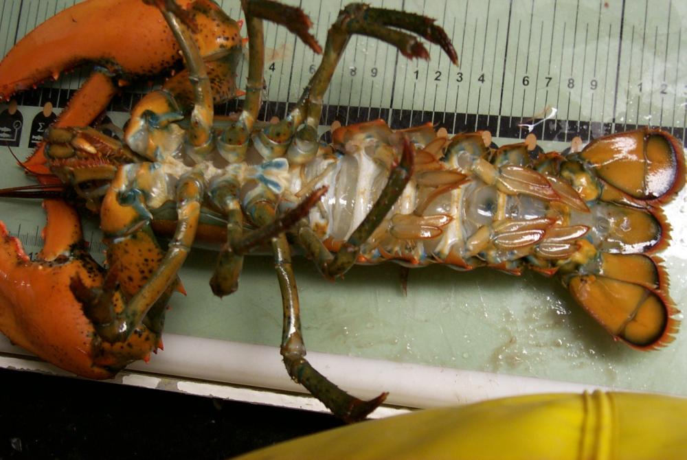
Figure 1. Ventral view of a lobster showing the clear intersegmental
cuticle. Color of the hemolymph could be observed non-invasively
through theis cuticle and that color checked by subsequent bleeding.
This specimen was photographed on Leg IV of Albatross IV 2002 Spring Groundfish
Survey by JK Kunkel.
My current hypothesis about serum and ovary color is somewhat changed
from previous hypotheses. A female who is beginning to deposit vitellogenin
from the blood into developing eggs has a light orange hemolymph that is
often seen as clear hemolymph through the cuticle. Its ovaries may
be orange to yellowish-green to green but the ovary is small in diameter
(0.5 cm at most. The hemolymph is orange because production of Vg
by the hepatopancreas is ahead of uptake into the ovaries and thus the
orange Vg can be seen in the serum. When ovaries are maximally growing
the hemolymph is in generally clear because the ovaries are efficiently
clearing Vg from the hemolymph. When the eggs are chorionating the
uptake of Vg stops and some Vg piles up in the serum and the serum may
become dark orange. The ovaries of dark orange serum females are
always large in diameter with visible eggs within the ovarian epithelium.
The one female with green hemolymph had flaccid large ovaries which I suspect
were involuting. There were no signs of ovulated eggs in this female.
The green hemolymph females are most often found in pounds and in caged
captive females and I continue to believe these are not healthy reproducing
females. Analysis of the hemolymph and ovarian samples this summer
will allow a better assessment of this current hypothesis. Each frozen
ovary will be examined to measure the diameter of the developing oocytes
and the status of enzymatic processing of its Vg peptides.
-
Work on a collaboration to study Lobster Shell Disease
A collaboration with Dr. Diane Cowan to extend our interest in the normal
serum patters of lobsters to shell disease is developing. I talked
with a DVM, Stephen Ellis, employed by Maine Department of Marine Resources
working on the aquacultured salmon diseases. He was sampling fish
caught on the Albatross while in the northern Gulf of Maine for the viral
disease organisms that is currently attacking Maine salmon pens.
He told me of a shell disease outbreak in Maine waters which occurred several
years ago and discussed various scenarios that might have contributed to
the outbreak. While that outbreak has passed, the current outbreak
spreading from Long Island Sound is possibly related and some greater collaboration
with the Maine lobster experience should be explored. The collaboration
with Diane Cowan becomes more important and sampling in both the Massachusetts
area as well as the central Gulf of Maine lobster areas is warranted.
Personnel
Dr. Diane Cowan. President of The Lobster Conservency collaborated in
providing lobsters that could be bled at regular intervals.
Jeff Xu applied his skills at database management improving an online
database of Lobster serum collection information.
Dennis Carrol, a UMass undergraduate has worked in the lab as a lab
technician, mainly helping to keep the lab glassware and working spaces
orderly. He has also learned to take images of specimens using our
image acquisition system. His current project will be to photograph
the ovary samples and catalog the lobster serum specimens brought back
from the AL02-03 leg IV cruise.