Origin of Lesions

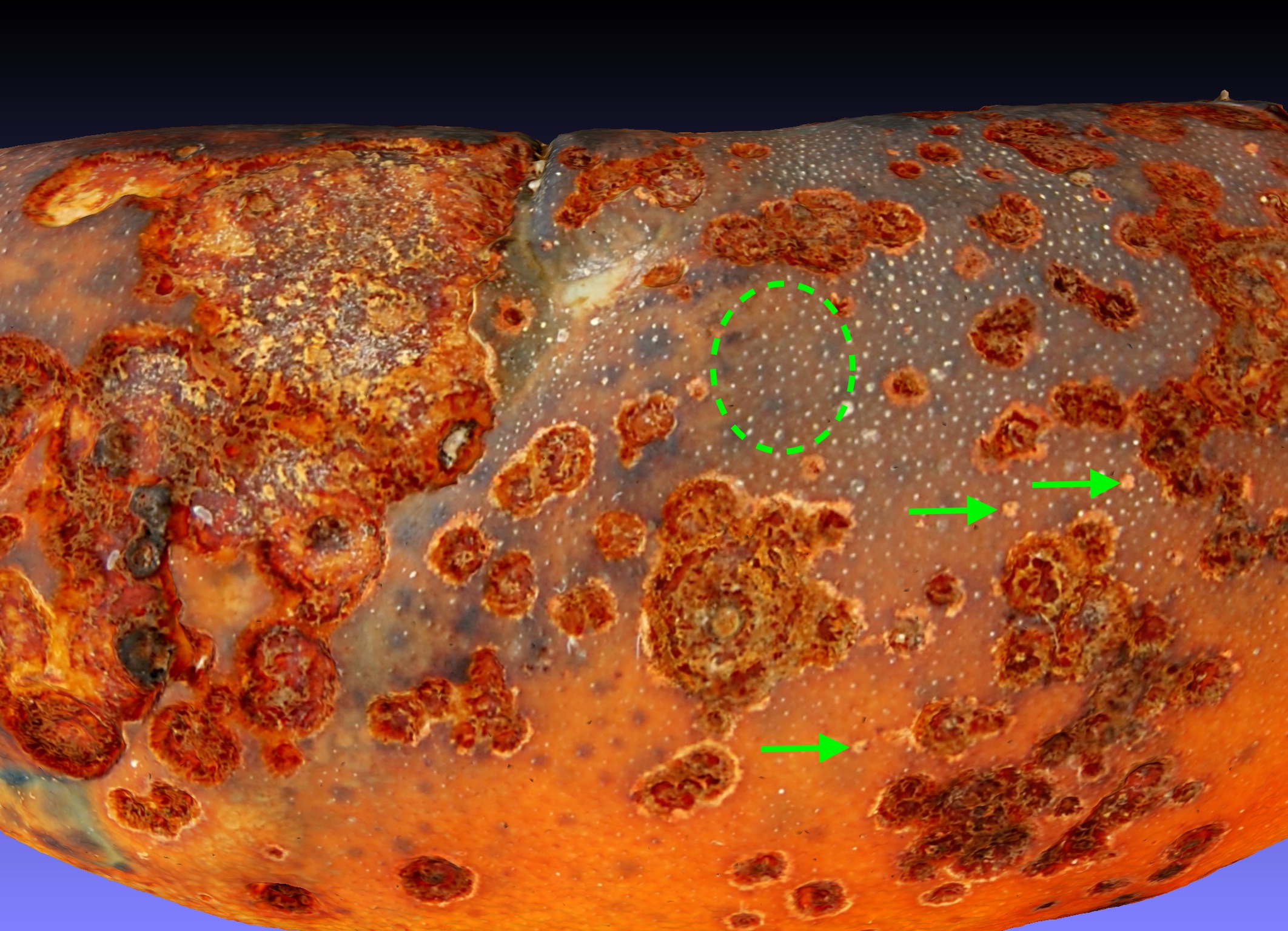
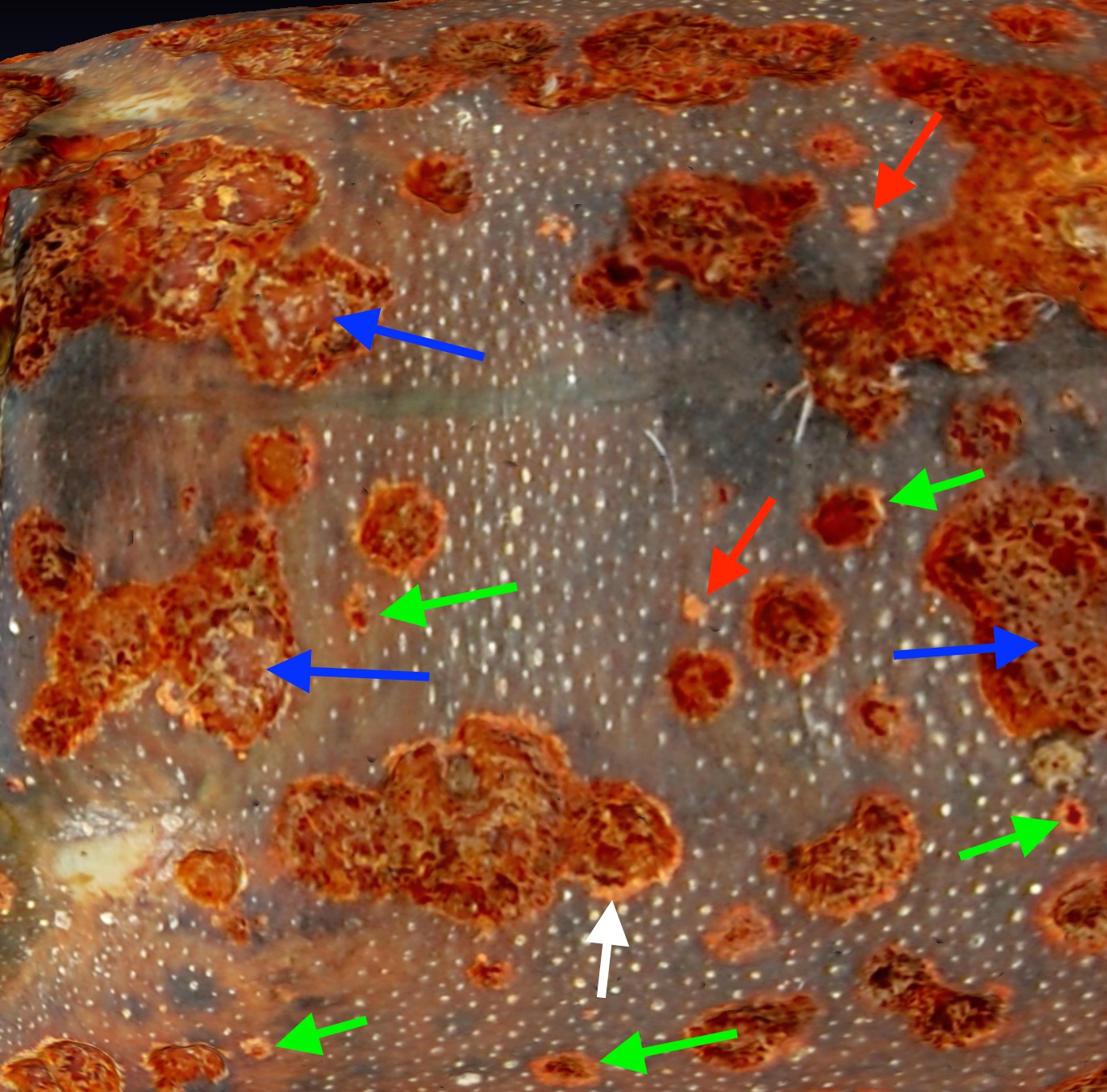
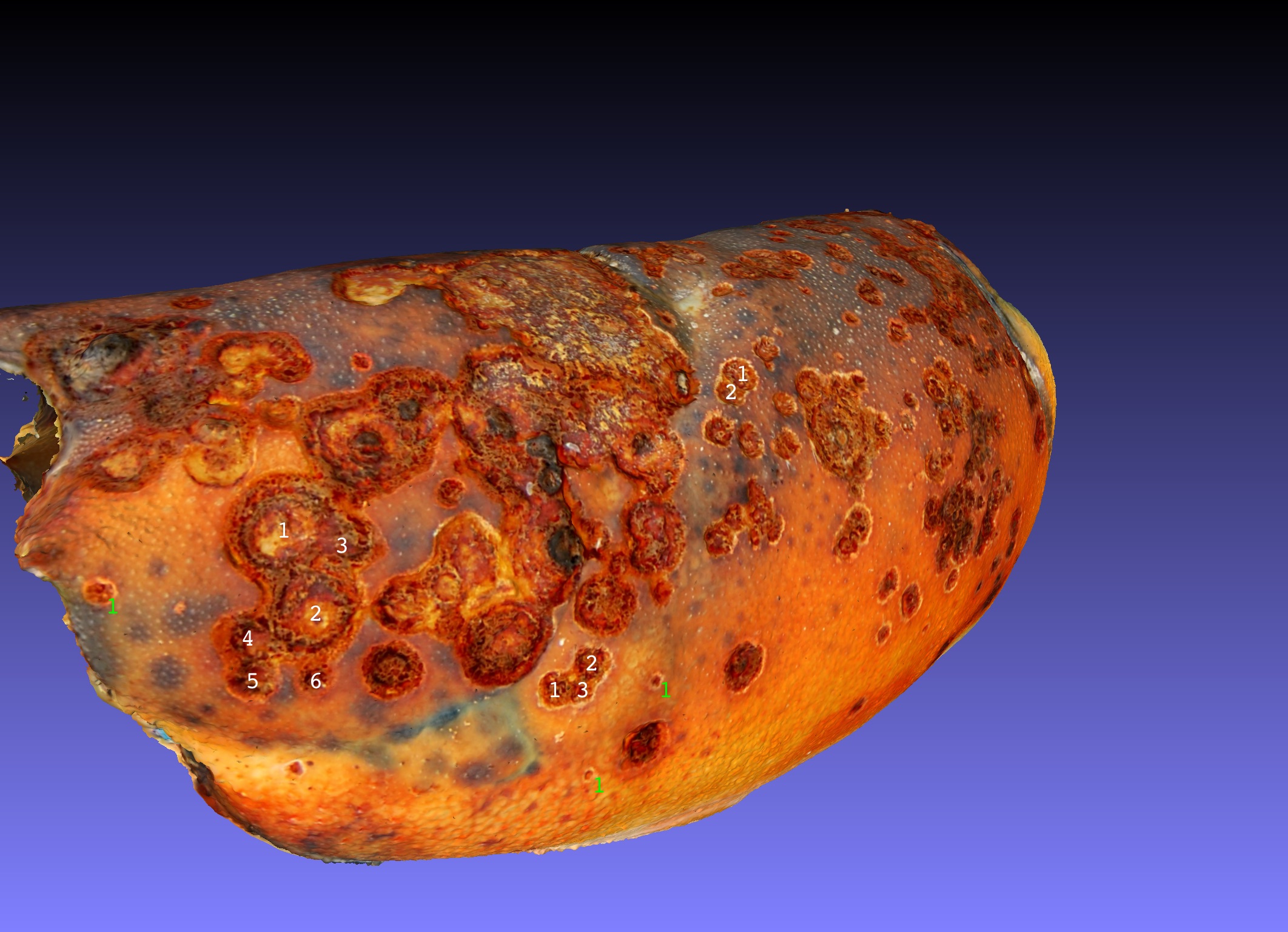
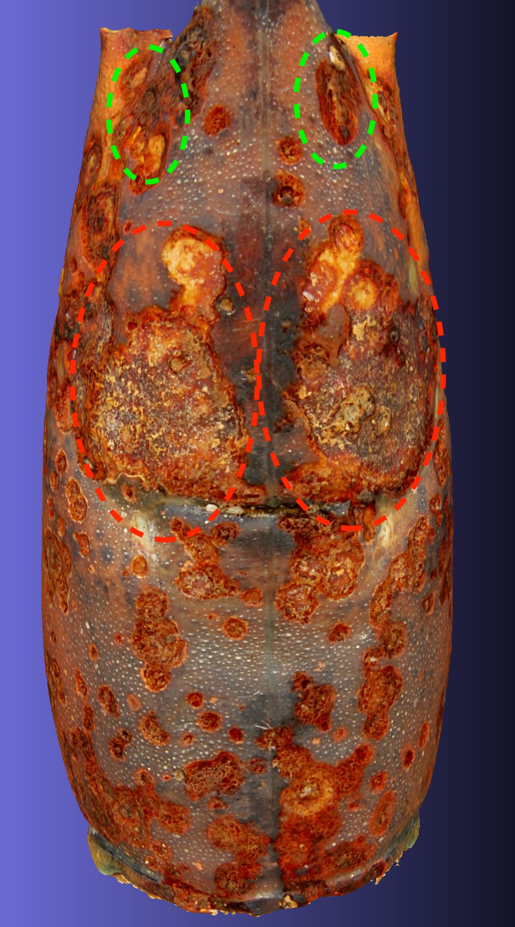
The origin of lobster shell lesions has been discussed particularly after the importance of Epizootic Shell Disease (ESD) became apparent. A description of its pathology and that of related diseases has been discussed (Chistoserdov et al. 2005). In the accompanying figure I show a sector of the S2019_398_01 carapace which exhibits level 3 ESD and shows what I interpret as origins that occured most recently pointed to by green arrows. The green dashed oval surrounds a field of cuticle that is populated by primary, secondary and tertiary organules that have been discussed as potential origins of lesion attack.

Development of Lesions

The development of lobster shell lesions in ESD is displayed in the accompanying figure. I show a sector of the S2019_398_01 carapace in which 3 stages of ESD are shown. The red arrows point to the initial stage of lesion development which is erosion in the calcite layer of the shell. It is primarily calcite that is light brown colored. The green arrows point to the second stage of lesion development which enters the exocuticle and in which a melanization of the lesion occurs, seen as a dark brown center inside the light brown calcite layer. Finally the blue arrows point to stage three of lesion development which reaches into the endocuticle in which the matrix is lighter than the melanized exocuticle with irregular structure that may correspond to the palisades of degraded cuticle polymer including melanin accumulated during the lesion enlargement.

Lesions Merge

The merger of lobster ESD lesions is demonstrated in the accompanying figure in which I show a sector of the S2019_398_01 carapace which exhibits 3 examples of ESD lesions. The green '1's point to single lesions. Clusters of lesions that have merged are marked by white number series 1-2, 1-3, and 1-6. Other clusters of lesions are so advanced that the original outlines of the merged lesions are obscured.

Symmetry of Lesions

The ESD lobster shell lesions displayed in the accompanying figure are largely symetrical in distribution about the dorsal midline of the carapace. The dorsal midline suture itself is largely devoid of lesions. Lesions of ESD typically develop first about the dorsal anterior midline of the carapace. As lesions are initiated and accumulate they merge and a pattern of distribution is often evident. In many cases there are approximately symetrical clusters of lesions on left and right side of the carapace as seen in the accompanying figure. The red dashed ovals surround major aggregations of lesions in the mid-dorsal region. A green dashed pair of ovals urround another aggregation of lesions in the anterior dorsal carapace. This symetrical distributions of lesions was foretold by symetrical distribution of early lesions that eventually merged. The pattern of symetry is repeated in other ESD infected carapaces:

Bibliography